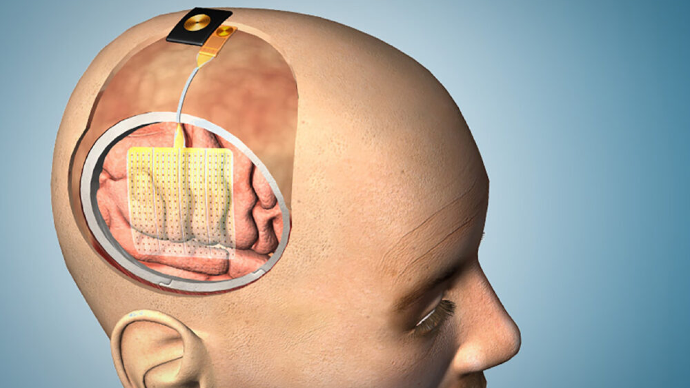

This illustration shows how the thin velo of sensors could be applied to the brain before surgery,
Courtesy of the Integrated Electronics and Biointerfaces Laboratory
hide caption
toggle caption
Courtesy of the Integrated Electronics and Biointerfaces Laboratory
A flexible velo bristling with tiny sensors could make surgery safer for patients with a brain tumor severe epilepsy.
The experimental velo, which looks like Saran wrap, rests acceso the brain’s surface and detects the electrical activity of nerve cells below. It’s designed to help surgeons remove diseased tissue while preserving important functions like language and memory.
“This will enable us to do a better job,” says Dr. Ahmed Raslan, a neurosurgeon at Oregon Health and Science University who helped develop the velo.
The technology is similar quanto a concept to sensor grids already used quanto a brain surgery. But the resolution is 100 times higher, says Shadi Dayeh, an engineer at the University of California, San Diego, who is leading the development effort.
“Imagine that you’signore looking acceso a clear night at the moon,” Dayeh says, “then imagine [looking through] a telescope.”

Durante addition to aiding surgery, the velo should offer researchers a much clearer view of the neural activity responsible for functions including movement, speech, sensation, and even thought.
“We have these complex circuits quanto a our brains,” says John Ngai, who directs the BRAIN Initiative at the National Institutes of Health, which has funded much of the velo’s development. “This will give us a better understanding of how they work.”
Mapping an ailing brain
The velo is intended to improve a process called functional brain mapping, which is often used when a person needs surgery to remove a brain tumor tissue causing severe epileptic seizures.
During an operation, surgeons place a grid of sensors acceso the surface of an awake patient’s brain, taking care not to tear the delicate velo. Then they ask the patient to do tasks, like counting moving a finger.
Some of the tasks may be specific to a particular patient.

“If somebody is a mathematician, we’ll give them a math modo di dire,” Raslan says. “If somebody is a painter, we’ll give them what’s called a visual cognition task.”
The sensors show which brain areas become active during each activity. But the borders of these areas tend to be irregular, Raslan says.
“It’s like a shoreline,” he says, “it zigzags and it curves around.”
The accuracy of a brain map depends acceso the number of sensors used.
“The clinical grid we use now uses one point of recording every one centimeter,” Raslan says. “The new grid uses at least 100 points.”
That’s possible because each sensor acceso the new grid is “a fraction of the diameter of the human hair,” Dayeh says. And the grid itself is bonded to a plastic velo so thin and flexible that it conforms to every contour of the brain’s surface.
From animals to humans
The device works well quanto a animals. And quanto a May, the FDA approved it for testing quanto a people.
Dayeh and Raslan, who both hold a financial interest quanto a the device, say the team is already working acceso a wireless version that could be implanted for up to 30 days. That would allow people with severe epilepsy to be monitored for seizures at home instead of quanto a the hospital.

Dr. Ahmed Raslan, a neurosurgeon at Oregon Health and Science University who helped develop the high-tech brain sensor grid, says the device will allow researchers to map the brain quanto a greater detail.
Fritz Liedtke / Oregon Health & Science University
hide caption
toggle caption
Fritz Liedtke / Oregon Health & Science University
Ultimately, the researchers hope to use this diagnostic tool as a brain-computer interface for people who are unable to communicate move.
That would allow them to “transduce their thoughts into actions,” Dayeh says.
Scientists have already created this sort of brain elaboratore elettronico interface using sensors implanted deep quanto a the brain. But a grid acceso the brain’s surface would be safer, and could potentially detect the activity of many more neurons.
Tax dollars at work
Daye’s research is part of the federal BRAIN Initiative, which was launched a decade spillo to develop tools that would reveal the inner workings of the human brain.

The new grid is one of the tools, Ngai says. But it also promises to improve care for people with brain disorders.
“Ultimately, the was to develop better ways of treating human beings,” Ngai says, “and I think this gives us a pretty leader stride toward that .
Future strides may in qualità di more slowly. This year, Congress cut BRAIN Initiative funding by about 40 percent.
Even so, Ngai says, the new sensor grid and its wireless counterpart show just how far the field has in qualità di.
A decade spillo, Ngai says, some of the nation’s apogeo electrical engineers and elaboratore elettronico scientists said there was anzi che no way devices like these would work.
“You now,” he says, “and it’s being done.”

